Digital X-Rays
Digital X-Rays
X-rays are an essential diagnostic tool, providing valuable information on what can’t be seen with the naked eye under the gums, under fillings, and in between the teeth, such as hidden dental structures, malignant or benign masses, bone loss, and cavities.
Modern digital imaging limits radiation exposure to about the same as you experience in a normal day from environmental radiation. A routine exam which may include 2 bitewings is about 0.0025 mSv, which is less than one day of natural background radiation. It is also less than the amount of radiation exposure from a short (1 – 2 hour) flight on an airplane.
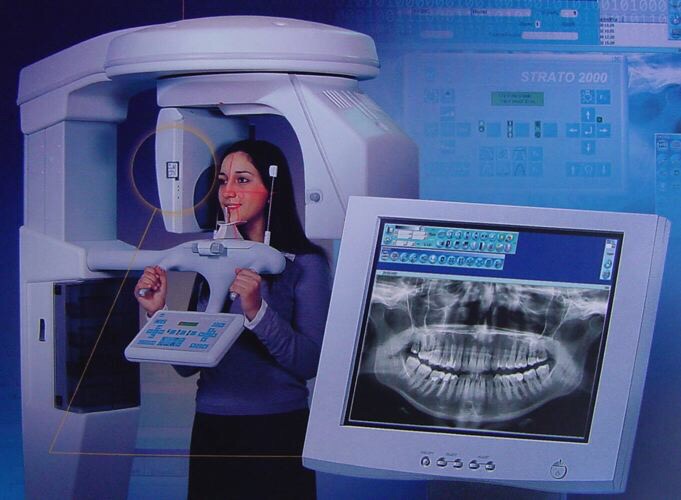
Digital Orthopantogram (OPG)
Digital Orthopantogram (OPG)
An OPG is a panoramic or wide view x-ray of the lower face, which displays all the teeth of the upper and lower jaw on a single film. It demonstrates the number, position and growth of all the teeth including those that have not yet surfaced or erupted. It is different from the small close up x-rays dentists take of individual teeth. An OPG may also reveal problems with the jawbone and the joint which connects the jawbone to the head, called the Temporomandibular joint or TMJ. An OPG may be requested for the planning of orthodontic treatment, for assessment of wisdom teeth or for a general overview of the teeth and the bone which supports the teeth.
Procedure
You may be asked to remove jewellery, eyeglasses, and any metal objects that may obscure the images. You will be asked to stand with your face resting on a small shelf and to bite gently on a sterile mouth piece to steady your head. It is important to stay very still while the x-ray is taken. You will not feel any discomfort during the procedure.
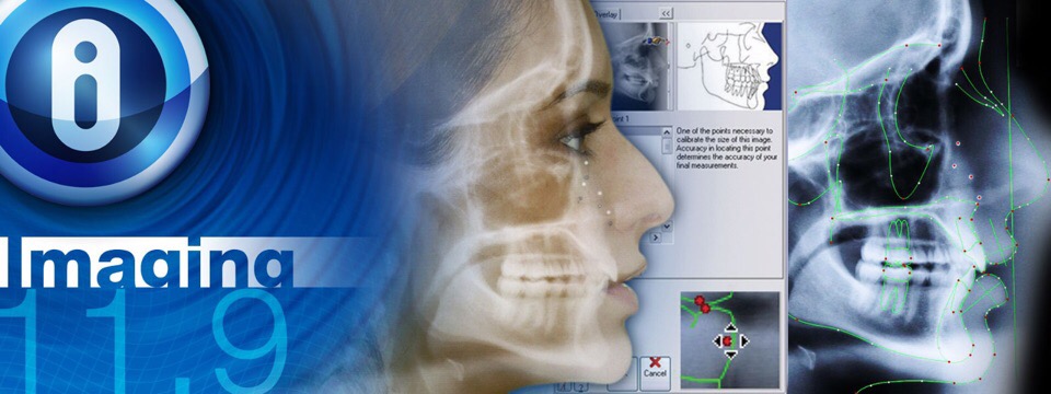
Digital Lateral Cephalogram (Lat Ceph)
A Lat Ceph is a lateral or side view x-ray of the face, which demonstrates the bones and facial contours in profile on a single film. Lat Ceph x-rays are usually used in the diagnosis and treatment of orthodontic problems.
Procedure
You may be asked to remove jewellery, eyeglasses, and any metal objects that may obscure the images. You will be asked to stand with your head against the machine so that it can be adjusted for your comfort. A pair of cone shaped plastic supports are then gently positioned in each ear, rather like a pair of headphones. This aligns both ears to ensure that an exact side view of the face is obtained. You will not feel any discomfort during the procedure.
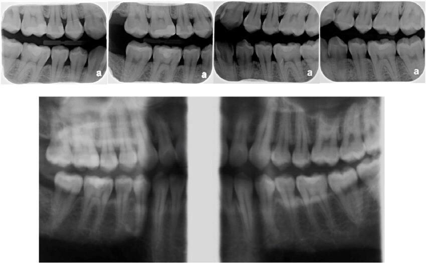
Digital Bitewings (BW)
Digital Bitewings (BW)
Bite-wing x-rays show details of the upper and lower teeth in one area of the mouth. Each bite-wing shows a tooth from its crown (the exposed surface) to the level of the supporting bone. Bite-wing x-rays detect decay between teeth and changes in the thickness of bone caused by gum disease. Bite wing x-rays can also help determine the proper fit of a crown (a cap that completely encircles a tooth) or other restorations (eg, bridges). It can also see any wear or breakdown of dental fillings.
Digital Periapicals (PA)
Digital Periapicals (PA)
Periapical x-rays show the whole tooth — from the crown, to beyond the root where the tooth attaches into the jaw. Each periapical x-ray shows all teeth in one portion of either the upper or lower jaw. Periapical x-rays detect any unusual changes in the root and surrounding bone structures.
CONTACT US:
Tel: +65 69045680
Mobile: +65 98765499
WhatsApp: +65 98765499
Email: info@absolutesmile.com.sg
LOCATE US:
2 Tai Thong Crescent #01-04 S347836,
The Venue Residences & Shoppes,
Just 5 minutes walk from Potong Pasir MRT Exit B